Galería de imágenes
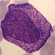
Cell description:
Size: 15-25 µm
Nucleus: oval with identifiable nucleoli and diffuse chromatin structure Cytoplasm: basophilic with visible golgi-zone and eye-catching azurophil granula (primary granulation)
<p>Cell description: </p> <p>Size: 15-25 µm </p> <p>Nucleus: oval with identifiable nucleoli and diffuse chromatin structure Cytoplasm: basophilic with visible golgi-zone and eye-catching azurophil granula (primary granulation)</p>
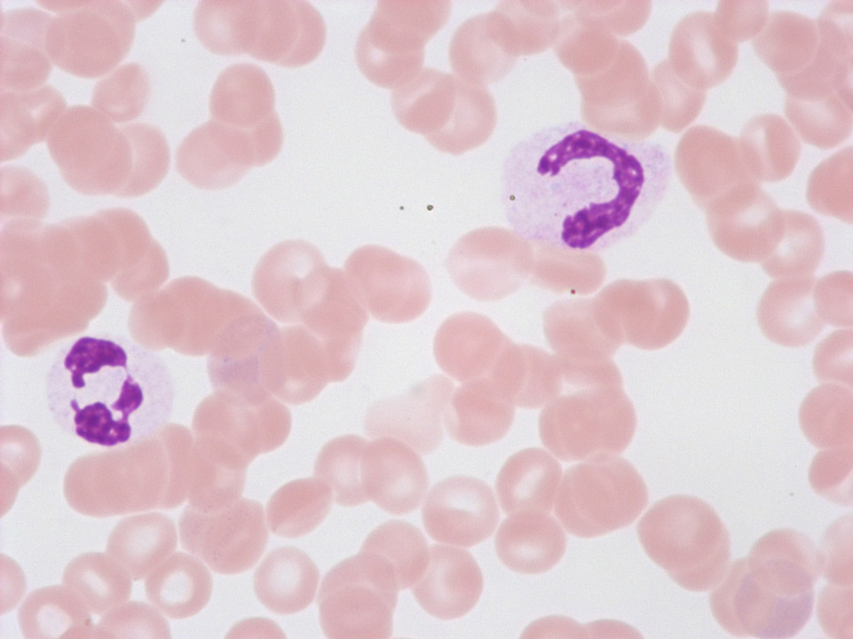
These so-called 'pseudo-Pelger's nuclear anomalies' with abnormal chromatin condensation and/or deficient nuclear segmentation are often found with viral infections, after chemotherapy, or with a myelodysplastic syndrome (MDS). Conversely, the true Pelger's nuclear anomaly is hereditary and without pathological significance.
<p>These so-called 'pseudo-Pelger's nuclear anomalies' with abnormal chromatin condensation and/or deficient nuclear segmentation are often found with viral infections, after chemotherapy, or with a myelodysplastic syndrome (MDS). Conversely, the true Pelger's nuclear anomaly is hereditary and without pathological significance.</p>
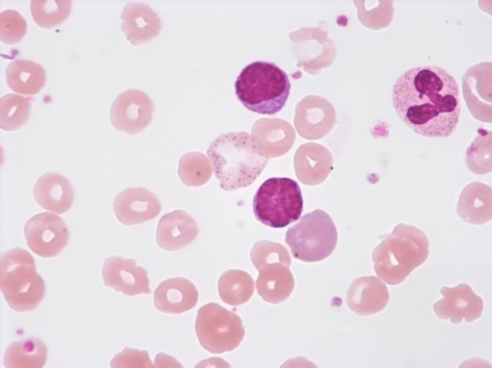
Large red blood cell with basophilic stippling (between the two lymphocytes) in a case of auto-immune haemolytic anaemia (AIHA). To the right, next to the lower lymphocyte, there is a polychromatic red blood cell.
<p>Large red blood cell with basophilic stippling (between the two lymphocytes) in a case of auto-immune haemolytic anaemia (AIHA). To the right, next to the lower lymphocyte, there is a polychromatic red blood cell.</p>
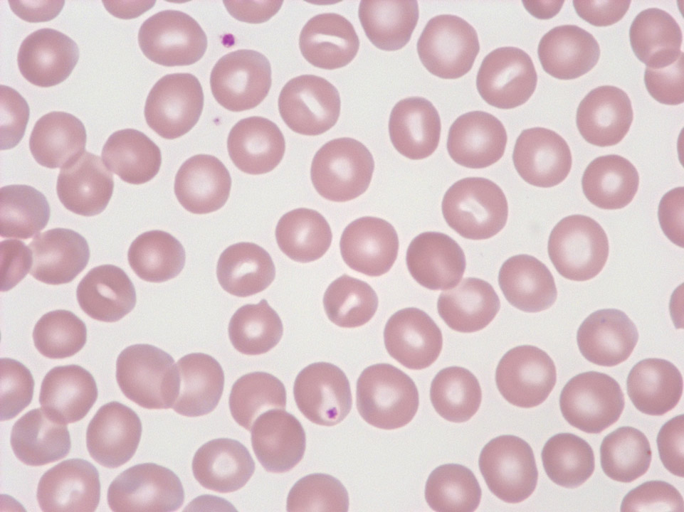
Red blood cells of a healthy individual showing a paler centre and a darker border. Occasionally the pale centre has an elongated shape (stomatocytes; at this frequency they have no pathological significance).
<p>Red blood cells of a healthy individual showing a paler centre and a darker border. Occasionally the pale centre has an elongated shape (stomatocytes; at this frequency they have no pathological significance).</p>
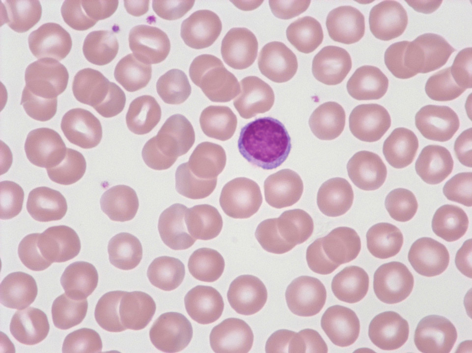
Red blood cells of a healthy individual. Normal red blood cells are a little smaller than the 'standard lymphocyte', which is pictured here.
<p>Red blood cells of a healthy individual. Normal red blood cells are a little smaller than the 'standard lymphocyte', which is pictured here.</p>
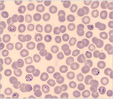
Trophozoites (rings) of Plasmodium falciparum are often thin and delicate, measuring on average 1/5 the diameter of the red blood cell. Rings may possess one or two chromatin dots. They may be found on the periphery of the RBC and multiple-infected RBC are not uncommon. Ring forms may become compact or pleomorphic, depending on the quality of the blood or if there is a delay in making smears.
<p>Trophozoites (rings) of Plasmodium falciparum are often thin and delicate, measuring on average 1/5 the diameter of the red blood cell. Rings may possess one or two chromatin dots. They may be found on the periphery of the RBC and multiple-infected RBC are not uncommon. Ring forms may become compact or pleomorphic, depending on the quality of the blood or if there is a delay in making smears. </p>
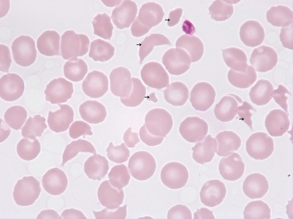
Schistocytes (->) and thrombocytopenia in a patient with haemolytic uraemic syndrome (HUS). This disease, as well as for example thrombotic thrombocytopenic purpura (TTP), is a so-called 'microangiopathic haemolytic anaemia' (MAHA). Characteristic of MAHA is a responsive increase in the production of red blood cells and platelets whose immature precursors (reticulocytes and immature platelet fraction, IPF) can be measured on certain Sysmex analysers. On a blood film the differential diagnosis of MAHA is not possible.
<p>Schistocytes (->) and thrombocytopenia in a patient with haemolytic uraemic syndrome (HUS). This disease, as well as for example thrombotic thrombocytopenic purpura (TTP), is a so-called 'microangiopathic haemolytic anaemia' (MAHA). Characteristic of MAHA is a responsive increase in the production of red blood cells and platelets whose immature precursors (reticulocytes and immature platelet fraction, IPF) can be measured on certain Sysmex analysers. On a blood film the differential diagnosis of MAHA is not possible.</p>
