Galería de imágenes
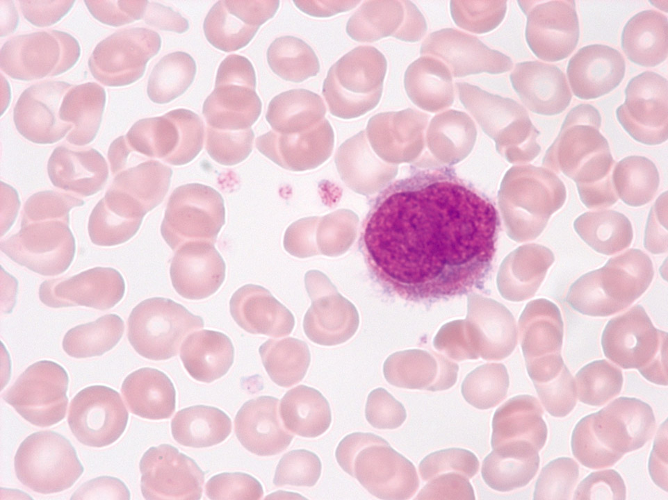
In primary myelofibrosis (PMF, also called chronic idiopathic myelofibrosis, CIMF) nuclei of megakaryocytes can be detected in the peripheral blood (May-Grünwald-Giemsa stain) and rarely, like here, small megakaryocytes. Both derive from extramedullary haematopoiesis.
<p>In primary myelofibrosis (PMF, also called chronic idiopathic myelofibrosis, CIMF) nuclei of megakaryocytes can be detected in the peripheral blood (May-Grünwald-Giemsa stain) and rarely, like here, small megakaryocytes. Both derive from extramedullary haematopoiesis.</p>
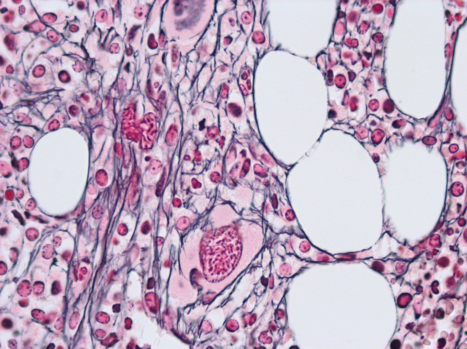
In the bone marrow histology the reticular fibres in PMF (alo called CIMF) can be identified by their black colour (Gomori stain).
<p>In the bone marrow histology the reticular fibres in PMF (alo called CIMF) can be identified by their black colour (Gomori stain). </p>
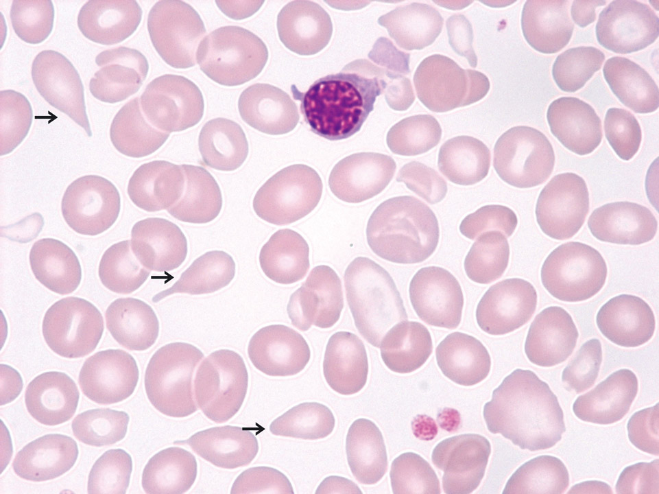
PMF (also called CIMF) in the fibrotic stage with a leucoerythroblastic blood picture (May-Grünwald-Giemsa stain): In this patient immature granulocytes, erythroblasts and many teardrop cells (->) were found. In the fibrotic stage there is usually anaemia present and a low to normal or reduced platelet count. Myeloblasts might be present. But more than 10% already indicate blast cell excess or transition into AML.
<p>PMF (also called CIMF) in the fibrotic stage with a leucoerythroblastic blood picture (May-Grünwald-Giemsa stain): In this patient immature granulocytes, erythroblasts and many teardrop cells (->) were found. In the fibrotic stage there is usually anaemia present and a low to normal or reduced platelet count. Myeloblasts might be present. But more than 10% already indicate blast cell excess or transition into AML. </p>
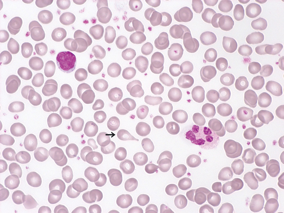
This peripheral blood film (May-Grünwald-Giemsa stain) is from a patient with PMF (also called CIMF) in the prefibrotic stage. You can see scattered teardrop cells (->). Furthermore, a few granulocytic precursors and very few erythroblasts were detected (not shown). The automated cell count showed slight leukocytosis, thrombocytosis and anaemia.
<p>This peripheral blood film (May-Grünwald-Giemsa stain) is from a patient with PMF (also called CIMF) in the prefibrotic stage. You can see scattered teardrop cells (->). Furthermore, a few granulocytic precursors and very few erythroblasts were detected (not shown). The automated cell count showed slight leukocytosis, thrombocytosis and anaemia.</p>

Cell description:
Size: 10-18 µm, smaller than lymphoblast
Nucleus: round with coarser structure than a lymphoblast, one distinct nucleolus
Cytoplasm: blue without granules
<p>Cell description: </p> <p>Size: 10-18 µm, smaller than lymphoblast </p> <p>Nucleus: round with coarser structure than a lymphoblast, one distinct nucleolus </p> <p> Cytoplasm: blue without granules </p>
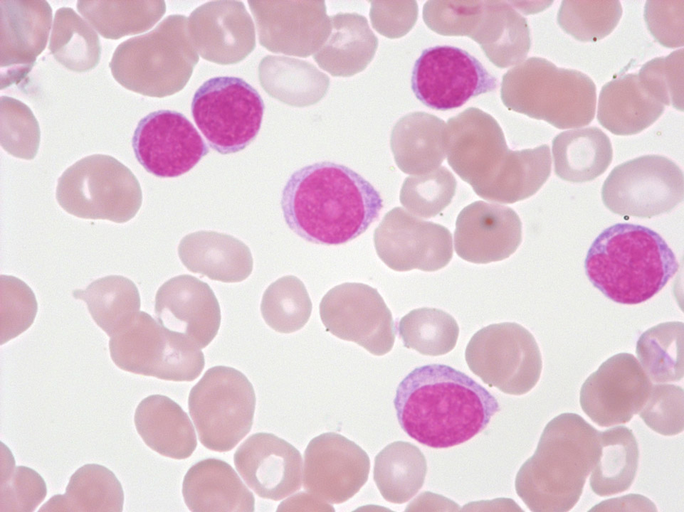
EDTA blood of a female patient with a transitional form of chronic lymphocytic leukaemia/prolymphocytic leukaemia. The three large cells are prolymphocytes, the smaller ones are lymphocytes.
<p>EDTA blood of a female patient with a transitional form of chronic lymphocytic leukaemia/prolymphocytic leukaemia. The three large cells are prolymphocytes, the smaller ones are lymphocytes.</p>
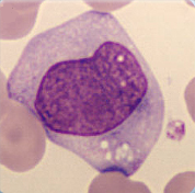
Cell description:
Size: bigger than monoblast
Nucleus: oval, kidney-shaped or lobulated, diffuse chromatin pattern, sometimes with nucleoli
Cytoplasm: pale basophil with fine azurophil granula.
Cell division is still possible.
<p>Cell description: </p> <p>Size: bigger than monoblast </p> <p>Nucleus: oval, kidney-shaped or lobulated, diffuse chromatin pattern, sometimes with nucleoli </p> <p>Cytoplasm: pale basophil with fine azurophil granula. </p> <p>Cell division is still possible.</p>
