Galería de imágenes

Cell description:
Size: up to 20 µm
Nucleus: eccentric with coarsely clumped chromatin, often clock-face chromatin pattern
Cytoplasm: strongly basophilic cytoplasm with apparent less basophilic Golgi zone adjacent to the nucleus
<p>Cell description: </p> <p>Size: up to 20 µm </p> <p>Nucleus: eccentric with coarsely clumped chromatin, often clock-face chromatin pattern </p> <p>Cytoplasm: strongly basophilic cytoplasm with apparent less basophilic Golgi zone adjacent to the nucleus</p>

Bone marrow histology (Giemsa stain) of a patient with multiple myeloma showing groups (nests) of plasma cells (->) in atypical locations. Normal plasma cells are usually located close to the blood vessels.
<p>Bone marrow histology (Giemsa stain) of a patient with multiple myeloma showing groups (nests) of plasma cells (->) in atypical locations. Normal plasma cells are usually located close to the blood vessels.</p>

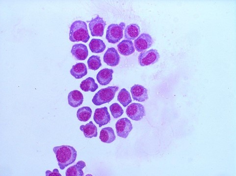
Plasma cells in a cytospin prepared from cerebrospinal fluid (CSF) of the patient with multiple myeloma, May-Gruenwald Giemsa stain
<p>Plasma cells in a cytospin prepared from cerebrospinal fluid (CSF) of the patient with multiple myeloma, May-Gruenwald Giemsa stain</p>

It is of utmost importance to always report a correct value for the platelet concentration. When detecting thrombocytopenia the laboratory should therefore double-check that it is not due to platelet aggregates (->) (pseudo-thrombocytopenia). In a haematology analyser they are usually automatically identified and flagged. In a blood film they are mostly located at the feather edge or the lateral edges.
<p>It is of utmost importance to always report a correct value for the platelet concentration. When detecting thrombocytopenia the laboratory should therefore double-check that it is not due to platelet aggregates (->) (pseudo-thrombocytopenia). In a haematology analyser they are usually automatically identified and flagged. In a blood film they are mostly located at the feather edge or the lateral edges.</p>

Platelets can lie on top of red blood cells, and look very much like Plasmodia. (By turning the micrometre screw, one will realize quite easily that the object is on top of the red blood cell and not inside it, as it would be with Plasmodia.)
<p>Platelets can lie on top of red blood cells, and look very much like Plasmodia. (By turning the micrometre screw, one will realize quite easily that the object is on top of the red blood cell and not inside it, as it would be with Plasmodia.)</p>

Platelet satellitosis is a rare phenomenon of platelets aggregating around polymorphonuclear white blood cells. It appears to be induced or enhanced by the presence of EDTA, a commonly used anticoagulant, and is not associated with any definite disease process.
<p>Platelet satellitosis is a rare phenomenon of platelets aggregating around polymorphonuclear white blood cells. It appears to be induced or enhanced by the presence of EDTA, a commonly used anticoagulant, and is not associated with any definite disease process.</p>

